Home
Home PG College
 Patient Login
 Sancheti Clinic
 Contact Us
 Download Reports

Sciatica Explained: Causes, Symptoms, and Effective Treatments
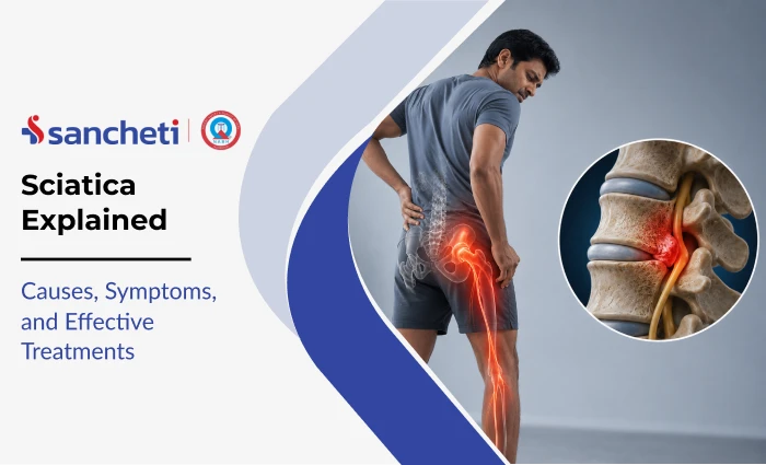
Sciatica is pain that radiates along the path of the sciatic nerve, from the lower back through the buttock and down one leg, sometimes reaching the foot. It is not a standalone diagnosis but a symptom of an underlying condition that compresses the sciatic nerve or its spinal nerve roots.
In the large majority of cases, sciatica resolves with structured conservative management, and surgery is not required.
At Sancheti Hospital, Pune, our Spine Department manages the full spectrum of sciatic nerve pain to provide relief, from acute disc herniation causing sudden severe leg pain, to chronic spinal stenosis producing progressive walking difficulty in older patients. This article explains the causes, symptoms, diagnostic approach, and treatment options clearly and in full.
What Is Sciatica?
The sciatic nerve is the longest and widest nerve in the human body. It originates from five nerve roots in the lumbar and sacral spine, which merge in the pelvis to form the sciatic nerve. From there it passes through the buttock, behind the hip joint, and down the back of the thigh to the lower leg and foot.
When any of these nerve roots are compressed at the point where they exit the spine, pain, tingling, and numbness follow the nerve’s anatomical path. This is what produces the characteristic single-leg symptoms of sciatica: pain that starts in the lower back or buttock and travels downward.
The medical term for sciatica caused by spinal nerve root compression is radiculopathy (from the Latin radix, meaning nerve root). The distinction matters clinically, because the level of compression determines which dermatome (skin area) and myotome (muscle group) are affected, and therefore guides both diagnosis and treatment.
Who It Affects
Sciatica has a lifetime prevalence of 13-40% of the general population, with an annual incidence of 1-5% (StatPearls, NIH). Peak incidence occurs in the fourth decade of life, and there is no significant gender predominance. The following risk factors have been consistently identified across multiple studies:
| Risk Factor | Evidence |
|---|---|
| Age 40-60 years | Peak incidence in the fourth decade; risk rises with disc degeneration |
| Obesity | Increases risk of hospitalisation for sciatica |
| Smoking | Hazard ratio 1.49 for sciatica hospitalisation |
| Heavy lifting at work | Hazard ratio 2.10 for sciatica hospitalisation; most consistent occupational risk |
| Sedentary work with awkward loads | Hazard ratio 1.57 vs lighter sedentary work |
| Whole-body vibration exposure | Particularly high risk when combined with obesity (HR 3.50 for obese exposed workers) |
| Physical inactivity | Walking or cycling to work reduces hospitalisation risk by 33% |
| Pre-existing spinal degeneration | Narrows foramina, increasing the likelihood of nerve root compression |
A 2024 study over 15 years found that older age, low education, psychological symptoms, multimorbidity, overweight, obesity, physical inactivity, and current smoking were all positively associated with sciatica. The majority of identified risk factors are modifiable, meaning targeted prevention is possible.
Sciatica Causes
Sciatica has a specific anatomical cause in the vast majority of cases. Identifying that cause, and not simply treating the pain pattern, determines the correct treatment pathway. The same symptom (radiating leg pain) can arise from several different structural problems, each requiring a different approach.
| Cause | Mechanism | Who It Affects Most |
|---|---|---|
| Lumbar disc herniation | Nucleus pulposus (gel centre of a disc) protrudes and presses directly on a nerve root | Adults 30-50 years; most common cause overall |
| Lumbar spinal stenosis | Spinal canal or foramina narrow, compressing nerve roots — often from bone spurs and ligament thickening | Adults over 60; the leading cause in older patients |
| Spondylolisthesis | One vertebra slips forward over the one below, narrowing the foramen through which the nerve root exits | Any age; degenerative or isthmic (from stress fracture) |
| Piriformis syndrome | Piriformis muscle in the pelvis compresses the sciatic nerve directly, not at spinal level | Runners, cyclists, and those with prolonged sitting; often misdiagnosed |
| Degenerative disc disease | Loss of disc height reduces foramen size, narrowing the space available for nerve roots | Adults over 40 with cumulative disc wear |
| Pregnancy | Enlarged uterus and postural shifts compress sciatic nerve in the third trimester | Third trimester; typically resolves post-delivery |
Piriformis syndrome accounts for 6-8% of patients presenting with sciatic-distribution pain (Comprehensive Spine Care). It is frequently overlooked because the pain pattern is indistinguishable from spinal-level sciatica in history alone. The distinction requires clinical examination and, in some cases, imaging, and treatment differs significantly from disc-related sciatica.
Symptoms of Sciatica
The symptom pattern of sciatica is clinically distinctive. The key feature is unilateral leg pain that follows a dermatomal (nerve distribution) path, not generalised or bilateral back pain, which suggests a different diagnosis.
| Symptom | Clinical Detail |
|---|---|
| Unilateral radiating leg pain | Almost always one leg; bilateral symptoms suggest spinal stenosis or serious pathology |
| Pain from lower back through buttock and down the leg | Follows the sciatic nerve path; may reach the calf, ankle, or foot |
| Burning, electric, or shooting quality | Distinct from the dull ache of muscular pain; indicates nerve irritation |
| Tingling or pins-and-needles in the leg or foot | Sensory nerve fibre involvement; location indicates which root is compressed |
| Numbness in the leg or foot | More advanced nerve compression; the specific area numb correlates to the nerve root level |
| Weakness in the leg or foot | Motor nerve fibre involvement; indicates significant compression; needs prompt assessment |
| Pain worse with prolonged sitting | Disc pressure increases in flexion, aggravating the compressed root |
| Pain worse with coughing or sneezing | Momentary rises in spinal pressure aggravate the compressed nerve |
In disc-related sciatica, walking or standing often provides relief because spinal extension reduces disc pressure on the nerve root. In spinal stenosis, the opposite is often true, patients find relief by leaning forward or sitting (flexion reduces the degree of narrowing). This distinction can help clinicians differentiate the underlying cause before imaging is performed.
Diagnosis for Sciatica
Accurate diagnosis requires identifying both the symptom pattern and its anatomical source. Sciatica from a disc herniation produces a different pattern of numbness and weakness than sciatica from piriformis syndrome, and each requires a different treatment approach.
Clinical Examination
- Straight Leg Raise (SLR) test: The patient lies flat and the examiner raises the extended leg. A positive test reproduces pain radiating below the knee at 30-70 degrees of elevation. It is the most sensitive clinical test for lumbar nerve root compression from disc herniation.
- Motor, sensory, and reflex testing: Examining specific muscle groups (myotomes), skin areas (dermatomes), and deep tendon reflexes allows the clinician to map which nerve root is affected before imaging. Loss of the ankle reflex, for example, points to S1 involvement.
Imaging and Investigations
- MRI: The gold standard for sciatica investigation. MRI directly visualises disc herniations, spinal canal narrowing, nerve root compression, and soft tissue pathology. It is required before any surgical decision and when red flag symptoms are present.
- X-ray: Identifies bony alignment problems, disc space narrowing, bone spurs, and vertebral slippage (spondylolisthesis). Does not visualise disc or nerve tissue directly.
- Nerve Conduction Study (NCS) and EMG: Confirms nerve root involvement, identifies which root is affected, and grades the severity of nerve dysfunction. Essential before surgery when clinical and imaging findings need correlation.
- CT scan: Used where MRI is contraindicated (pacemaker, certain implants) or where bony detail of foraminal stenosis is needed in addition to soft tissue information.
How Sciatica is Treated
Conservative Sciatica treatment leads to up to 90% improvement in disc-related sciatica within 4 months (NEJM, 2019). For these reasons, the treatment pathway begins conservatively and advances to interventional options only if adequate improvement does not occur.
Step 1: Activity Management and Analgesia (Weeks 1-2)
Bed rest is not recommended. Remaining gently active, walking, moving around the house, is consistently superior to immobilisation for sciatica recovery. Prolonged sitting and sustained spinal flexion should be avoided.
- NSAIDs (anti-inflammatory medications) for pain and nerve inflammation
- Neuropathic agents (gabapentin, pregabalin) for burning or electric-quality leg pain
- Short-term muscle relaxants if significant paraspinal muscle spasm is present
- Ice or heat application to the lower back for symptomatic relief
Step 2: Physiotherapy and Exercise (Weeks 2-12)
A structured programme overseen by a physiotherapist is the cornerstone of sciatica recovery. Active exercise outperforms passive modalities consistently in clinical evidence.
- Neural mobilisation (nerve flossing/gliding): Gentle sciatic nerve mobilisation exercises reduce nerve adhesion, improve nerve mobility, and decrease radicular pain
- McKenzie extension exercises: For disc-related sciatica, repeated lumbar extension often centralises leg pain (moves it from the foot towards the back), indicating disc repositioning and nerve root decompression
- Core stabilisation: Strengthening the deep lumbar stabilisers (transverse abdominis, multifidus) reduces mechanical load on the affected disc and nerve root
- Walking programme: Graduated daily walking improves spinal circulation, maintains mobility, and supports recovery across all causes of sciatica
Step 3: Epidural Steroid Injection (Weeks 4-12 if Inadequate Progress)
Epidural steroid injection (ESI) delivers corticosteroid directly into the epidural space adjacent to the compressed nerve root, reducing local inflammation. ESI is most useful as a bridge, it reduces pain sufficiently to allow participation in physiotherapy and return to activity.
Ultrasound-guided or fluoroscopy-guided injection is recommended over landmark-based injection for improved accuracy and outcomes.
Step 4: Surgery (Indicated Sciatica Not Resolving after 6-12 Weeks, or Urgent Red Flags)
Surgery is not the default for sciatica. It is appropriate when:
- Symptoms are severe and have not responded to structured conservative treatment for 6-12 weeks
- Progressive neurological deficit (increasing weakness, expanding area of numbness)
- Cauda equina syndrome, emergency surgery within 24-48 hours
- Patient preference after being fully informed of equivalent long-term outcomes with conservative management
Surgical Options:
- Microdiscectomy: The standard procedure for disc herniation causing sciatica. A small incision (2-3 cm) is made and the portion of disc compressing the nerve root is removed under microscopic vision.
- Laminotomy / Endoscopic Decompression: For spinal stenosis, the bony and ligamentous structures narrowing the spinal canal are partially removed to relieve nerve root pressure
- Spinal fusion (where indicated): Reserved for cases with significant instability, spondylolisthesis, or recurrent disc herniation requiring structural stabilisation
Recovery From Sciatica
Recovery from sciatica depends on the underlying cause, the severity of nerve compression, and how long symptoms have been present before treatment begins. The nerve itself determines the pace of improvement: once compression is relieved, whether by conservative treatment or surgery, the nerve heals at its own biological rate.
Conservative Recovery Timeline
| Timeframe | Expected Progress |
|---|---|
| Week 1-2 | Active pain management; gentle mobility; avoid bed rest and prolonged sitting |
| Weeks 2-6 | Physiotherapy and nerve mobilisation begin; leg pain starts to centralise (move towards back) |
| Weeks 6-12 | Strengthening and functional rehabilitation; most patients return to work |
| Months 3-6 | Full recovery for most acute cases; leg symptoms typically resolve last |
| Months 6-12 | Persistent or chronic sciatica cases; nerve regeneration continues gradually |
Post-Surgery Recovery Timeline
| Procedure | Hospital Stay | Return to Light Work | Return to Physical Work |
|---|---|---|---|
| Microdiscectomy | 1-2 days | 2-4 weeks | 6-12 weeks |
| Laminotomy / Endoscopic Decompression | 1-3 days | 4-6 weeks | 10-14 weeks |
| Spinal Fusion | 3-5 days | 6-8 weeks | 3-6 months |
Rehabilitation Phases
Whether recovering conservatively or post-surgery, a structured physiotherapy and rehabilitation programme is essential for restoring full function and preventing recurrence.
Phase 1: Pain Control and Neural Calm (Weeks 1-2)
- Avoid bed rest; remain gently mobile to maintain spinal circulation
- Positions of relief: walking or lying with knees slightly bent, not prolonged sitting
- Ice or heat to the lower back for symptom management
- Medication as prescribed; neuropathic agents for burning leg pain
Phase 2: Neural Mobilisation and Flexibility (Weeks 2-6)
- Sciatic nerve flossing (neural mobilisation), gentle leg movements that mobilise the nerve within its sheath and reduce adhesion to surrounding tissue
- McKenzie extension programme for disc-related sciatica; reduces disc pressure and centralises leg pain
- Progressive walking programme, the single most accessible and effective recovery activity
- Hamstring and hip flexor stretching, both muscle groups increase neural tension in the sciatic nerve when tight
Phase 3: Core Stabilisation and Strengthening (Weeks 6-12)
- Deep core activation (transverse abdominis, multifidus) to offload the affected spinal segment
- Hip strengthening, the gluteal muscles are frequently weakened in sciatica and need targeted rehabilitation
- Graduated return to work activities, with ergonomic assessment if occupational factors contributed to the episode
- Progressive endurance training to restore cardiovascular fitness lost during the acute phase
Phase 4: Return to Full Activity (Weeks 12 onwards)
- Sport-specific or occupation-specific training reintroduced under physiotherapist guidance
- Long-term maintenance programme; core and hip strengthening, flexibility, postural habits
- Monitoring for recurrence; early intervention at first signs of symptom return
Prevention of Sciatica
Most risk factors for sciatica are modifiable. Targeted lifestyle changes reduce both the likelihood of a first episode and the risk of recurrence after recovery.
- Maintain a healthy weight: Obesity increases sciatica hospitalisation risk. Even modest weight loss reduces spinal load significantly.
- Correct lifting technique: Lift with the legs, not the back; keep the load close to the body; avoid twisting while holding weight. Frequent lifting carries a 2.1-fold increased risk of sciatica hospitalisation
- Walk or cycle to work: Active commuting reduces sciatica hospitalisation risk by 33%, independent of body weight and other leisure activities (PubMed, 2017).
- Avoid prolonged sitting: Stand up and move at least every 30-45 minutes; use a sit-stand workstation if available; keep hips and spine in neutral position
- Core strengthening: Strong deep core muscles (transverse abdominis, multifidus) protect lumbar discs from the mechanical forces of daily activity and sport
- Stop smoking: Smoking impairs blood flow to intervertebral discs, accelerating degeneration and increasing sciatica risk. Smoking carries a hazard ratio of 1.49 for sciatica hospitalisation
- Act on early symptoms: Intermittent leg tingling or back pain that appears on exertion responds well to conservative management. Waiting until symptoms are constant and muscles are weakening substantially worsens outcomes
Key Takeaways
- Sciatica is a symptom of underlying nerve compression, not a standalone diagnosis. Identifying the cause determines the correct treatment.
- Lifetime prevalence is 13-40%; annual incidence 1-5%; peak in the fourth decade. The majority of risk factors are modifiable.
- The most common cause is lumbar disc herniation. In patients over 60, spinal stenosis becomes the leading cause. Piriformis syndrome is frequently misdiagnosed.
- Most of the patients recover without surgery. Conservative treatment leads to major improvement in disc-related sciatica within 4 months.
- Red flag symptoms: bladder or bowel dysfunction, saddle numbness, bilateral leg weakness; indicate potential cauda equina syndrome, a surgical emergency requiring immediate hospital attendance.
- Epidural steroid injection surpasses other conservative methods short-term but does not provide long-term results; it is most effective as a bridge to active physiotherapy.
- Surgery (microdiscectomy) speeds short-term recovery but produces equivalent long-term outcomes to conservative management in non-emergency cases. It is a last resort after 6-12 weeks of structured conservative care.
- Obesity, smoking, heavy lifting, and physical inactivity are the main modifiable risk factors. Walking or cycling to work reduces sciatica hospitalisation risk by 33%.
- At Sancheti Hospital, Pune, our Spine Department and physiotherapy team evaluate the cause, severity, and functional goals of each patient before recommending any treatment, ensuring the right intervention, at the right time.
Frequently Asked Questions (FAQs)
Q1. How do I know if my back and leg pain is sciatica or something else?
The hallmark of sciatica is unilateral radiating leg pain that follows a dermatomal path, from the lower back through the buttock and down the leg, often with tingling, numbness, or weakness. Non-specific back pain that stays in the back without radiating below the buttock is not sciatica. Hip pain that radiates into the groin rather than down the leg usually originates from the hip joint, not the spine. The positive straight leg raise test, pain below the knee when the extended leg is raised, is the most reliable clinical differentiator. An MRI in Pune confirms the diagnosis and identifies the cause.
Q2. Can sciatica go away on its own without treatment?
In many cases, yes, particularly when caused by disc herniation. The natural history of sciatica from lumbar disc herniation is favourable: 60-80% of patients see significant improvement within 6-12 weeks. The herniated disc material is often gradually reabsorbed by the body over weeks to months. However, waiting without treatment is not the same as doing nothing: staying active, avoiding aggravating positions, and beginning specific exercises significantly speeds recovery and reduces the likelihood of symptoms persisting beyond 3 months.
Q3. Should I rest in bed or stay active with sciatica?
Stay active. This is one of the most consistently supported recommendations in sciatica management. Prolonged bed rest worsens outcomes, it does not reduce nerve root inflammation and leads to deconditioning, increased pain sensitivity, and delayed recovery. The goal in the first 1-2 weeks is activity modification: avoid sustained spinal flexion (prolonged sitting, bending forward) and high-impact loading. Gentle walking is beneficial from the beginning. A targeted physiotherapy programme, started within the first 2 weeks, consistently outperforms passive rest in clinical studies.
Q4. How long does sciatica last?
For acute disc-related sciatica, 75% of patients see meaningful improvement within 4 weeks. Up to 20-30% of patients develop symptoms that persist beyond 3 months, defined as chronic sciatica, and these cases require a more structured treatment approach including physiotherapy, possible epidural injection, and surgical evaluation if there is no improvement. Chronic sciatica (lasting over 6 months) that requires surgery has poorer surgical outcomes than early-stage sciatica, which is one reason why identifying cases that are not responding to conservative treatment at 6-8 weeks, and acting on them, matters clinically.
Q5. When does sciatica need surgery?
Surgery is indicated in four situations: (1) cauda equina syndrome or any red flag neurological emergency, surgery within 24-48 hours; (2) progressive motor weakness that is worsening despite conservative treatment; (3) severe sciatica that has not improved after 6-12 weeks of structured conservative management including physiotherapy and, where appropriate, epidural injection; and (4) patient preference after being fully counselled that surgical and conservative outcomes are equivalent by 12 months in non-emergency cases. Surgery is not the appropriate first step for acute sciatica without neurological emergency, regardless of pain severity.