Home
Home PG College
 Patient Login
 Sancheti Clinic
 Contact Us
 Download Reports
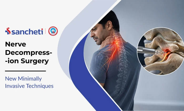

Nerve Decompression Surgery: New Minimally Invasive Techniques
Nerve decompression surgery is a procedure that relieves chronic pressure on a compressed nerve; eliminating pain, restoring function, and preventing permanent nerve damage.
When conservative measures such as physiotherapy, splinting, and steroid injections fail to resolve a pinched nerve, minimally invasive nerve surgery now offers patients faster recovery, smaller incisions, and significantly lower complication rates than traditional open surgery.
At Sancheti Hospital, Pune, our Hand and Microvascular Department and Spine Department treat the full range of nerve compression conditions; from carpal tunnel syndrome and cubital tunnel syndrome to lumbar disc herniation and spinal stenosis, using the latest minimally invasive techniques available.
What Is Nerve Decompression Surgery?
A pinched nerve occurs when surrounding tissue places sustained mechanical pressure on a nerve, disrupting its ability to transmit signals. The compressing structure may be a herniated disc, thickened ligament, narrowed bone channel, or scar tissue from a previous injury.
Nerve decompression surgery removes or repositions the structure causing compression. Once pressure is relieved, the nerve recovers. The critical variable is timing: delays in treatment allow progressive nerve damage that may not fully reverse even after a technically successful procedure.
Traditionally, nerve decompression required large open incisions, significant muscle stripping, and multi-day hospital stays. Advances in endoscopy, ultrasound guidance, and microsurgical instruments have fundamentally changed this. Most nerve decompression procedures are now performed through incisions measured in millimetres, under local or regional anaesthesia, as day-case procedures.
Who Needs Nerve Decompression Surgery?
Nerve decompression is indicated when a compressed nerve is:
- Causing constant (not merely intermittent) symptoms
- Failing to respond to 6-12 weeks of structured conservative treatment including physiotherapy and splinting
- Producing objective signs of nerve damage on electrodiagnostic testing
- Causing progressive muscle weakness or wasting that cannot wait for further conservative management
Conditions most commonly treated with nerve decompression surgery:
| Condition | Nerve Compressed | Location |
|---|---|---|
| Carpal tunnel syndrome | Median nerve | Wrist |
| Cubital tunnel syndrome | Ulnar nerve | Elbow |
| Radiculopathy (disc herniation) | Spinal nerve root | Cervical or lumbar spine |
| Spinal stenosis | Multiple nerve roots | Lumbar spine |
| Sciatica | Sciatic nerve root | Lumbar spine |
| Thoracic outlet syndrome | Brachial plexus | Neck-shoulder junction |
| Tarsal tunnel syndrome | Tibial nerve | Ankle |
Carpal tunnel syndrome (CTS) is the most prevalent compression neuropathy, affecting approximately 5% of the general population worldwide (Musculoskeletal Care, 2024). Cubital tunnel syndrome; compression of the ulnar nerve at the elbow is the second most common, with an estimated annual incidence of 25 per 100,000 people (ScienceDirect, 2021).
Grades of Nerve Compression
The severity of nerve compression determines whether surgery is required and how urgently. Grading is based on clinical examination and electrodiagnostic findings:
| Grade | Clinical Features | Typical Management |
|---|---|---|
| Mild | Intermittent tingling; symptoms provoked by position or activity; no muscle weakness | Conservative: splinting, physiotherapy, activity modification |
| Moderate | Frequent or constant tingling; some sensory loss; early grip weakness | Conservative with close monitoring; surgery if no improvement at 6-12 weeks |
| Severe | Constant numbness; significant motor weakness; muscle wasting visible | Surgical decompression indicated; delay risks permanent nerve damage |
Critical clinical point: pain alone does not determine surgical urgency. Muscle weakness and wasting on examination, even in a patient whose pain is manageable, indicate significant motor nerve fibre loss and are often a more pressing indication for surgery than pain severity.
Symptoms That Indicate Surgery Is Needed
| Symptom | Clinical Significance |
|---|---|
| Constant numbness (not just at night) | Indicates sustained compression beyond intermittent pressure |
| Progressive muscle weakness | Nerve damage advancing; urgent assessment required |
| Thenar or hypothenar wasting | Chronic severe compression with motor fibre loss |
| Symptoms failing to improve at 3 months of conservative care | Strong indicator for surgical evaluation |
| Drop foot or weakness lifting the foot | Lumbar nerve root compromise; urgent assessment required |
| Bilateral symptoms | May indicate spinal canal pathology rather than peripheral entrapment |
Important clinical distinction: numbness that wakes the patient at night may still respond to conservative pinched nerve treatment. Numbness present throughout the day, regardless of activity or position, indicates a higher grade of compression that usually requires surgery.
Diagnosis Before Surgery
Accurate localisation of the compression point is the foundation of a successful nerve decompression. Decompressing the wrong anatomical level produces a failed outcome regardless of how well the surgery itself is performed.
The diagnostic pathway in Pune includes:
Nerve Conduction Study (NCS) and Electromyography (EMG): These tests measure nerve signal speed and strength, identify where transmission slows or fails, and grade the severity of compression. NCS and EMG are essential before any surgical decision for peripheral nerve entrapment to distinguish between mild, moderate, and severe compression.
MRI: The gold standard for spinal nerve compression. MRI visualises disc herniations, ligament thickening, bony canal narrowing, and the spatial relationship between each structure and the nerve roots it may be compressing.
High-Resolution Ultrasound: Increasingly used for peripheral nerve assessment. Ultrasound directly visualises nerve swelling at the compression point and guides surgical planning. For ultrasound-guided decompression procedures, pre-operative nerve mapping is a prerequisite.
X-ray: Rules out bony pathology, alignment problems, and calcification contributing to compression.
Treatment: Minimally Invasive Nerve Decompression Techniques
This is where surgical practice has advanced most dramatically. The choice of technique depends on which nerve is compressed, its anatomical location, the severity of compression, and the surgeon’s experience with each approach.
Endoscopic Carpal Tunnel Release
The most commonly performed endoscopic nerve decompression procedure. A portal incision of 1-2 cm is made at the wrist and a camera-equipped scope is introduced into the carpal tunnel. Under direct visualisation on a monitor, the transverse carpal ligament is divided with precision instruments.
This eliminates the large palm incision of traditional open carpal tunnel surgery and reduces scar tenderness, pillar pain (deep wrist pain after surgery), and return-to-work time.
Ultrasound-Guided Percutaneous Carpal Tunnel Release
The most recent innovation in carpal tunnel management. Under continuous high-frequency ultrasound imaging, a fine needle-based or thread-based device is passed through a skin opening under 5 mm. The transverse carpal ligament is divided with real-time visualisation of the median nerve, without any port or camera entering the carpal tunnel itself.
Endoscopic Cubital Tunnel Decompression (Ulnar Nerve)
Compression of the ulnar nerve at the elbow produces pain and tingling in the ring and little fingers, grip weakness, and in advanced cases, visible wasting of the small hand muscles. Endoscopic cubital tunnel release uses a single small incision at the elbow. A camera system visualises and decompresses the ulnar nerve along its course without the wide tissue dissection of open surgery.
When anterior transposition of the ulnar nerve is needed, this can also be performed under endoscopic guidance in experienced hands.
Microdiscectomy (Lumbar Nerve Root Decompression)
The minimally invasive standard for lumbar disc herniation causing sciatica. A small incision (2-3 cm) is made in the lower back. Using a surgical microscope, the portion of herniated disc material pressing on the nerve root is precisely removed. Muscle stripping is minimal compared to traditional open discectomy. Most patients mobilise within 24 hours and are discharged within 1-2 days.
For cervical disc herniation compressing neck nerve roots, anterior cervical discectomy or posterior cervical foraminotomy using tubular retractors achieves nerve decompression with similarly reduced tissue disruption.
Endoscopic and Tubular Laminotomy for Spinal Stenosis
Spinal stenosis, narrowing of the spinal canal that compresses multiple nerve roots, traditionally required laminectomy: a major open procedure stripping paraspinal muscles from both sides of the spine. Tubular retractor systems now allow decompression through a 16-22 mm working channel, dilating rather than stripping the muscles.
The older open-spine approach required 5-6 inch incisions and weeks of hospital stay; modern endoscopic procedures achieve equivalent decompression through openings of 2-5 mm.
Open Surgery vs. Minimally Invasive: Key Differences
| Factor | Traditional Open Surgery | Minimally Invasive Surgery |
|---|---|---|
| Incision size | 3-6 cm (peripheral) / 5-15 cm (spinal) | 2 mm to 3 cm |
| Anaesthesia | General (usually) | Local or regional (most cases) |
| Hospital stay | 1-5 days | Day case to 1-2 days |
| Blood loss | Higher | Minimal |
| Muscle handling | Significant stripping | Dilation, not stripping |
| Return to light work | 4-8 weeks | 1-3 weeks |
| Scar tenderness | More common | Minimal |
| Complication rate (cubital tunnel) | 12.9% | 7.9% |
Recovery After Nerve Decompression Surgery
The nerve itself determines the pace of recovery more than the surgical technique. Surgery removes the cause of compression; the nerve then heals at its own biological rate. The more severe and long-standing the compression before surgery, the slower and less complete the recovery.
Recovery Timeline
| Timeframe | What to Expect |
|---|---|
| Days 1-3 | Post-operative soreness at incision site; limb elevated; begin gentle active movement |
| Week 1-2 | Suture removal; grip and movement exercises begin; desk work possible for many patients |
| Weeks 2-6 | Progressive strengthening; sensation begins to improve; some tingling before resolution is normal |
| Weeks 6-12 | Return to most physical activities; muscle strength recovering |
| Months 3-6 | Full nerve recovery for moderate compression cases |
| Months 6-12 | Ongoing nerve regeneration in severe or long-standing compression cases |
Nerves do not heal in a straight line. Periods of rapid improvement followed by plateaus are entirely normal. Temporary worsening of tingling shortly after surgery, before improvement, is a common sign of nerve awakening, not deterioration.
Rehabilitation Phases
A structured physiotherapy and rehabilitation programme is essential in Pune for restoring full function after nerve decompression. Working with a qualified physiotherapist who specialises in post-surgical nerve recovery significantly improves outcomes.
Phase 1: Protection and Wound Healing (Days 1-14)
- Rest the operated limb; avoid loading the surgical site
- Elevation to control post-operative swelling
- Gentle active range-of-motion exercises to prevent stiffness
- Wound care and splinting where prescribed by the surgeon
Phase 2: Early Mobilisation and Nerve Gliding (Weeks 2-6)
- Nerve gliding exercises (also called nerve flossing) — controlled movements that mobilise the nerve within its tunnel and prevent re-adhesion to surrounding tissue
- Progressive grip and pinch strengthening for upper limb cases
- Walking programme for spinal decompression patients with gradual increase in duration and terrain
- Scar desensitisation to manage hypersensitivity at and around the incision
Phase 3: Strengthening and Functional Restoration (Weeks 6-12)
- Progressive resistance exercises targeting muscles weakened during the period of nerve compression
- Fine motor tasks for hand and wrist cases: typing, gripping, writing, turning small objects
- Core strengthening and back conditioning for spinal decompression patients
- Work-specific rehabilitation where occupational demands require targeted conditioning
Phase 4: Return to Full Activity (Weeks 12 onwards)
- Sport-specific or occupation-specific training reintroduced under physiotherapist guidance
- Return to driving, lifting, and strenuous manual tasks cleared by the surgeon
- Ongoing monitoring for incomplete nerve recovery or early signs of recurrence
Prevention
Not all nerve compression is preventable, but several modifiable factors significantly reduce the risk of reaching the stage where surgery is required:
- Ergonomic workstation setup: Keep wrists in a neutral position during keyboard use; position screens at eye level; support elbows to avoid prolonged flexion that places continuous stress on the ulnar nerve
- Avoid sustained pressure on nerves: Resting on elbows for long periods compresses the ulnar nerve; habitually crossing the knees can compress the peroneal nerve at the fibular head
- Maintain a healthy weight: Obesity is an established risk factor for carpal tunnel syndrome through increased canal pressure and systemic metabolic effects on nerve health
- Manage diabetes and hypothyroidism actively: Both conditions predispose to peripheral nerve compression and significantly impair nerve recovery after injury or surgery
- Act on symptoms early: Nerve entrapment diagnosed at the stage of intermittent symptoms responds well to conservative management. Waiting until numbness is constant and muscles are weakening substantially worsens surgical outcomes and reduces the likelihood of complete nerve recovery
Key Takeaways
- Nerve decompression surgery relieves pressure on a compressed nerve by removing or repositioning the compressing structure, eliminating pain, restoring nerve function, and preventing permanent damage.
- Surgery is indicated when symptoms are constant, muscles are weakening, or conservative treatment has produced no improvement after 6-12 weeks.
- Modern minimally invasive techniques: endoscopic release, ultrasound-guided percutaneous release, microdiscectomy, and tubular decompression, achieve outcomes equivalent to open surgery with significantly fewer complications, smaller incisions, and faster recovery.
- Nerve recovery after surgery is gradual and non-linear. Severity and duration of compression before surgery are the strongest predictors of how completely the nerve recovers.
- Structured physiotherapy is non-negotiable after nerve decompression, it prevents re-adhesion, rebuilds weakened muscles, and restores full functional use of the affected limb.
- At Sancheti Hospital, Pune, our Hand and Microvascular, Neurology, and Spine departments assess each patient’s nerve compression individually, selecting the surgical technique that best matches the nerve involved, its anatomical location, and the severity of damage.
Frequently Asked Questions (FAQs)
Q1. How do I know if I need nerve decompression surgery or if physiotherapy is enough?
The decision rests on three factors: severity of compression on electrodiagnostic testing, duration of symptoms, and whether conservative treatment is producing measurable improvement. Mild and moderate compression caught early with intermittent rather than constant symptoms, responds well to physiotherapy, splinting, and activity modification. When symptoms are constant, muscles are wasting, or objective nerve function tests are deteriorating despite adequate conservative management, surgery becomes the appropriate intervention. A specialist assessment with NCS and EMG is essential to make this distinction accurately.
Q2. Is minimally invasive nerve surgery as effective as traditional open surgery?
Yes, for the vast majority of cases. Systematic reviews comparing endoscopic and open carpal tunnel release show equivalent clinical outcomes and patient satisfaction scores. For cubital tunnel syndrome, the endoscopic technique delivers equivalent outcomes with a significantly lower complication rate. The advantage of minimally invasive approaches is not improved effectiveness; it is faster recovery, less post-operative pain, reduced scarring, and earlier return to work. In complex revision cases or where anatomy is unfavourable, open surgery may still be preferable for technical reasons.
Q3. How long after nerve decompression surgery will my numbness and tingling improve?
The timeline depends on how severely and for how long the nerve was compressed before surgery. Mild to moderate compression of relatively short duration often produces noticeable improvement within days to weeks. Severe or long-standing compression can take 6-12 months for meaningful nerve recovery, and where permanent nerve fibre loss has already occurred, complete reversal may not be possible. This is why early surgical intervention, when indicated, consistently produces better outcomes than delayed surgery.
Q4. Can nerve compression come back after surgery?
Recurrence is uncommon but possible. For carpal tunnel release, endoscopic techniques carry a recurrence rate of approximately 1.1% compared to 3.5% for open surgery in published meta-analyses (ScienceDirect, 2021). Recurrence is more likely when the underlying risk factors, repetitive hand or wrist loading, obesity, diabetes, are not addressed after surgery. A comprehensive post-surgical rehabilitation programme and ergonomic modifications significantly reduce the risk.
Q5. Will nerve decompression surgery be painful?
The surgery itself is performed under local or regional anaesthesia for most peripheral nerve cases, so the patient feels no pain during the procedure. Post-operative discomfort at the incision site is typically mild and managed with standard analgesics for 2-5 days. Many patients report that post-operative discomfort is substantially less than the chronic nerve pain they experienced before surgery. The more challenging aspect for most patients is not the surgical pain but the nerve recovery period; particularly when the nerve was severely compressed and is regenerating through the early phases of healing, during which temporary tingling is common and entirely normal.