Home
Home PG College
 Patient Login
 Sancheti Clinic
 Contact Us
 Download Reports
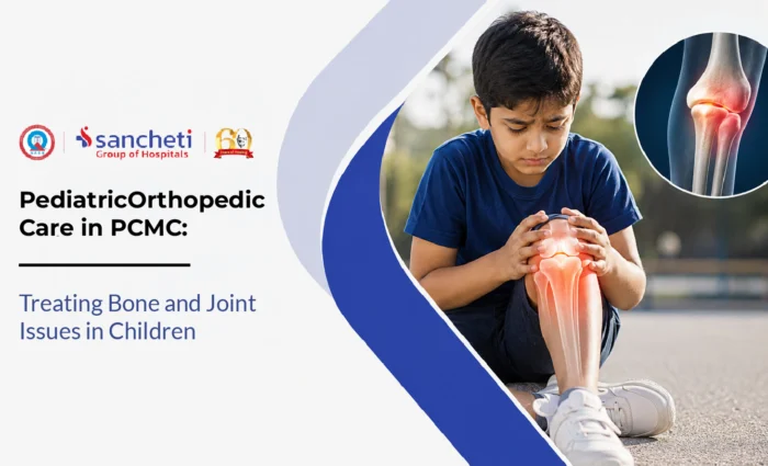

Paediatric Orthopaedic Care in PCMC: Treating Bone and Joint Issues in Children
Paediatric orthopaedic care is the branch of orthopedics that addresses bone, joint, and musculoskeletal conditions specifically in children and adolescents, from birth through the end of skeletal growth. It is a distinct clinical field from adult orthopedics because a growing skeleton behaves differently from a mature one.
For families across Pimpri-Chinchwad, Sancheti Hospital’s Paediatric Orthopaedic Department in Pune offers specialist care for the full range of childhood bone and joint conditions. This article helps parents understand which conditions need specialist attention, which are normal developmental variations, and what treatment involves at each stage of childhood.
What Is a Paediatric Orthopaedic Specialist?
A paediatric orthopaedic specialist is a surgeon with dedicated training in the musculoskeletal conditions of children, across every age from newborn to late adolescence. The distinction from a general orthopaedic surgeon matters in practice for several reasons.
A fracture through a growth plate in a six-year-old requires a different assessment, treatment, and follow-up protocol than a fracture in the same bone in a forty-year-old. A bow-legged two-year-old who is perfectly normal needs reassurance and observation; a bow-legged twelve-year-old needs investigation for underlying disease.
The paediatric orthopaedic doctor in PCMC also navigates the timing dimension that does not exist in adult care: using a growing skeleton’s remaining growth potential as a therapeutic tool, guiding bone growth toward correction in a child who still has years of development ahead, rather than performing an osteotomy (bone-cutting surgery) that will be needed if the window for growth modulation is missed.
Common Conditions Treated by a Paediatric Orthopaedic Specialist
A core task of the paediatric orthopaedic consultation in PCMC is distinguishing between conditions that genuinely need treatment and developmental variations that are normal for the child’s age and will resolve spontaneously.
| Condition | What It Is |
|---|---|
| Clubfoot (CTEV) | A congenital foot deformity in which the foot points downward and inward |
| Developmental dysplasia of the hip (DDH) | A spectrum of hip abnormality from mild laxity to full dislocation |
| Bow legs | An outward curve of the lower legs causing a gap between the knees when standing |
| Knock knees | An inward curve of the legs causing the knees to touch while the feet are apart |
| Adolescent idiopathic scoliosis (AIS) | A lateral curvature of the spine of 10 degrees or more, developing during the adolescent growth spurt |
| Growth plate fractures | Fractures through the growth plate (physis) at the end of long bones, which only exist in children |
| Legg-Calve-Perthes disease | Avascular necrosis (death of bone tissue due to disrupted blood supply) of the femoral head in children |
| Bone and joint infections | Bacterial infection of bone (osteomyelitis) or a joint (septic arthritis |
Symptoms That Warrant a Paediatric Orthopaedic Assessment
The clinical challenge in paediatric orthopedics is that many normal developmental milestones look worrying to parents, while some genuinely pathological findings produce no pain and are easy to miss.
| Symptom or Finding | Likely Significance |
|---|---|
| Visible foot deformity at birth | Possible clubfoot: refer to a paediatric orthopaedic specialist |
| A click, clunk, or limited hip movement | Possible DDH: requires urgent hip ultrasound |
| Limp without a history of injury | Possible Legg-Calve-Perthes disease or septic arthritis |
| A hot, swollen, painful joint with fever | Possible septic arthritis: orthopaedic emergency |
| One shoulder or hip visibly higher than the other | Possible scoliosis |
| Bow legs that are worsening rather than improving | Possible rickets or pathological genu varum |
| Persistent toe-walking beyond age 3 | May indicate tight Achilles tendon, neurological cause (cerebral palsy), or autism-related sensory pattern |
| Pain or swelling in a bone or joint that is constant | Possible bone tumour or infection |
| A child stops walking after previously walking, or regresses in motor milestones | Needs urgent assessment, both orthopaedic and neurological |
How Paediatric Orthopaedic Conditions Are Diagnosed
Diagnosis in paediatric orthopedics relies heavily on the developmental context, as the significance of any finding depends on the child’s age and expected developmental stage.
Clinical Assessment
The specialist will assess the child’s gait, posture, and range of motion, both in the affected area and overall.
Imaging by Age and Condition
- Ultrasound: the primary imaging tool for DDH in infants under four to six months; also used for soft tissue assessment in infections.
- X-ray: the primary tool for fractures, bone deformity assessment, scoliosis Cobb angle measurement, and Legg-Calve-Perthes staging.
- MRI: used for bone and joint infections where X-ray is normal but infection is suspected; for complex growth plate injuries; and for spinal cord assessment in scoliosis with atypical features.
- Blood tests: essential for suspected bone infections; for suspected rickets; and for juvenile idiopathic arthritis workup.
Observation Over Time
For many paediatric orthopaedic conditions, the most important diagnostic tool is serial observation at defined intervals. A single consultation cannot always provide a definitive answer; this follow-up process is part of the specialist care, not a limitation of it.
Treatment Options in Paediatric Orthopedics
Most paediatric orthopaedic conditions are treated conservatively, with surgery recommended only when it offers a clear long-term benefit. Many common concerns, such as in-toeing, flat feet, bow legs, or toe-walking in young children, are normal developmental variations that improve naturally with growth. In these cases, observation, regular follow-up, and reassurance are often all that is needed.
When treatment is required, it is tailored to the child’s age and condition. Congenital clubfoot is most commonly corrected using the Ponseti method, a series of gentle casts followed by bracing, with excellent success when started early. Braces are also used for conditions such as developmental hip dysplasia and scoliosis to support healthy growth and reduce the need for surgery. For significant bow legs or knock knees, guided growth surgery can gradually correct alignment while the child is still growing using a minimally invasive technique.
Surgery is reserved for conditions that cannot be corrected conservatively, such as severe deformities, displaced growth plate injuries, certain spinal curvatures, or complex bone and joint problems.
Recovery After Paediatric Orthopaedic Treatment
A growing child recovers from surgery faster than an adult in terms of bone healing, but conditions must be monitored until skeletal maturity.
| Treatment or Procedure | Typical Recovery and Follow-Up |
|---|---|
| Ponseti casting for clubfoot | Six to eight weeks of weekly casting, then brace worn full-time for three months reducing to nights and naps for four years. |
| Pavlik harness for DDH | Full-time harness use for six to twelve weeks, with regular ultrasound monitoring; gradual weaning as hip stability improves. |
| Guided growth surgery for angular deformity | Day-case procedure; normal walking resumes in two to three weeks; correction occurs progressively over six to eighteen months of bone growth. |
| Scoliosis surgery (posterior spinal fusion) | Four to five days in hospital; return to school in six weeks; back to most activities by three months. |
| Growth plate fracture (Salter-Harris types 3-4) | Cast immobilisation or surgical fixation followed by clinical and X-ray monitoring every three to four months for at least one year. |
| Septic arthritis drainage | Two to four days in hospital for intravenous antibiotics post-drainage; oral antibiotics for three to four weeks; physiotherapy to restore range of motion. |
Prevention and Early Detection
While not all paediatric orthopaedic conditions can be prevented, early detection and healthy habits can reduce the risk of serious problems.
- Newborn hip screening: Every baby should have hip examinations at birth and at the six-week check-up. Babies with risk factors, such as breech birth or a family history of hip problems, may also need an ultrasound.
- Early clubfoot treatment: Babies born with feet turned inward or downward should see a paediatric orthopaedic doctor in PCMC within the first week for the best results.
- Scoliosis checks: Parents of children aged 10–16 can perform the Adams forward bend test at home. A specialist should assess any visible asymmetry.
- Vitamin D: Adequate vitamin D supports healthy bone development, particularly in children with limited sun exposure.
- Maintain a healthy weight: Reduces stress on growing bones and joints during adolescence.
- Prevent sports injuries: Proper warm-ups, age-appropriate training, and avoiding overtraining help lower injury risk in active children.
Key Takeaways
- Children’s bones and joints are constantly growing, which means they require assessment and treatment strategies that differ significantly from those used in adults.
- Many concerns such as bow legs, knock knees, or flat feet are normal stages of development, making specialist evaluation important to distinguish harmless variations from conditions that require treatment.
- Early diagnosis allows many paediatric orthopaedic conditions to be managed with minimally invasive treatments that guide normal growth and reduce the need for surgery later in life.
- Prompt assessment of warning signs such as limping, persistent pain, visible deformities, or delayed motor milestones can prevent long-term complications and improve outcomes.
- Most paediatric orthopaedic conditions are managed conservatively through observation, casting, bracing, or guided growth, with surgery reserved for selected cases where it offers clear long-term benefit.
- Recovery in children is often faster than in adults, but regular follow-up remains essential to monitor bone growth and ensure normal development until skeletal maturity.
- Early screening, healthy bone development, injury prevention, and timely specialist care play a key role in supporting lifelong musculoskeletal health in children.
- For families across Pimpri, Chinchwad, Nigdi, Akurdi, Wakad, Bhosari, Tathawade, and Hinjewadi, Sancheti Hospital, Pune‘s Paediatric Orthopaedic Department, provides complete specialist care for children’s bone and joint conditions, from newborn assessment through to adolescent deformity management.
Frequently Asked Questions (FAQs)
Q1. My child walks with feet turned inward. Does this need treatment?
In-toeing is one of the most common reasons parents bring children to a paediatric orthopaedic specialist, and in the vast majority of cases, no treatment is needed. The three most common causes of in-toeing in young children are feet curved at birth, rotational alignment of the shin bone, and rotational alignment of the thigh bone. All three are developmental variants that correct spontaneously as the child grows, typically by age 8-10, without exercises, orthopaedic shoes, or corrective devices. The situations that warrant further assessment in Pune are: in-toeing in one leg only; in-toeing that is severe and affecting daily activities; or in-toeing associated with stumbling or falls that are progressing rather than improving.
Q2. My newborn has been diagnosed with clubfoot. What should we do immediately?
Act within the first week. Contact a paediatric orthopaedic specialist immediately for a Ponseti method assessment. The Ponseti method starts with gentle manipulation of the foot followed by a plaster cast from toes to thigh, changed weekly. Each cast gradually corrects the components of the deformity in a specific sequence. Most clubfoot cases require five to six casts over five to six weeks, followed by a minor Achilles tendon release as a day-case procedure, followed by a foot abduction brace. When treatment starts in the first two weeks of life, the neonatal tissue flexibility is at its maximum, and the correction achievable without surgery is at its greatest. This is one of the conditions where urgency of referral directly translates into quality of outcome.
Q3. My daughter’s shoulders look uneven. Could this be scoliosis, and how serious is it?
Uneven shoulders in an adolescent girl are a finding that warrants a specialist assessment, since adolescent idiopathic scoliosis has a strong female predominance and typically develops during the adolescent growth spurt. The appropriate first step is an Adams forward bend test: have her stand with feet together and bend forward with hands together, looking for an asymmetric rib hump or shoulder blade prominence as you view from behind. Any visible asymmetry on this test is a positive finding that justifies a specialist appointment. However, the severity of scoliosis varies enormously. Surgery for scoliosis is considered only for curves above 45-50 degrees or for curves that are documented to be progressing rapidly during active growth. The paediatric orthopaedic specialist appointment in Pimpri Chinchwad is for accurate measurement and monitoring, not necessarily for treatment.
Q4. My 5-year-old has flat feet. Do we need special shoes or insoles?
Probably not, and almost certainly not yet. Flat feet in children under six are entirely normal: the arch of the foot is largely composed of fat pad in early childhood, and the visible arch of an adult’s foot develops progressively as this fat pad redistributes. The muscles and ligaments of the arch strengthen. The foot of a healthy 5-year-old typically looks flat when standing, and this is a normal developmental stage, not a medical condition. The evidence for orthopaedic shoe inserts or arch-support insoles in asymptomatic childhood flat feet is consistently poor. Situations where flat feet do warrant specialist assessment are: flat feet that are rigid and do not form any arch even on tip-toe standing (flexible flat feet arch on tip-toe, rigid ones do not); flat feet associated with pain or difficulty walking; or flat feet that are significantly asymmetric.
Q5. How do I know if my child’s knee pain is growing pains or something that needs investigation?
Growing pains are a real and common phenomenon in children aged 3-12 years, characterised by aching pain deep in the muscles of the lower legs and thighs, typically occurring in the evenings and at night, affecting both legs, and resolving completely by the next morning without any physical findings on examination. The child walks normally; there is no swelling, no localised tenderness, and no pain during the day. Knee pain that warrants investigation is: pain that is present during the day and limits activity; pain associated with swelling of the joint; pain localised to a specific point around the knee rather than diffuse muscle aching; pain that is unilateral and constant; pain that wakes the child from sleep consistently; or pain associated with a limp. If your child’s joint pain persists, worsens, or affects walking or play, consult a kids joint pain doctor in Pimpri for a specialist assessment.