Home
Home PG College
 Patient Login
 Sancheti Clinic
 Contact Us
 Download Reports
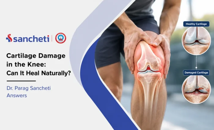

Cartilage Damage in the Knee: Can It Heal Naturally? Dr. Parag Sancheti Answers
One common question patients bring to a knee specialist is whether knee cartilage damage can heal on its own. The answer is more nuanced than a simple yes or no. Because articular cartilage has limited healing capacity, the outlook depends on the depth, size, and location of the damage, as well as the patient’s age and activity demands.
Dr. Parag Sancheti, Chairman and Managing Director of Sancheti Hospital and an internationally recognised knee surgeon with over 34 years of clinical and research experience, explains how cartilage damage is assessed, treated, and repaired through options that can realistically be achieved.
What Is Knee Cartilage and Why Does It Matter
Articular cartilage is the smooth, glistening tissue covering the ends of the bones inside the knee joint. It is approximately two to four millimetres thick and is composed of a specialised matrix of collagen fibres and proteoglycans maintained by cells called chondrocytes. It provides a low-friction, shock-absorbing surface that lets the knee move smoothly under load without the bone surfaces grinding against each other.
What makes articular cartilage biologically unusual is that it has no blood supply of its own. Chondrocytes receive their nutrition through diffusion from the surrounding synovial fluid rather than through blood vessels. No nerves run through the cartilage either, which helps explain why early cartilage damage may be painless until it reaches the underlying bone or becomes large enough to alter joint mechanics. This means significant defects can develop before symptoms become obvious.
Why Cartilage Cannot Heal Like Other Tissues
When a muscle tears or a bone breaks, blood drives the healing response. Injured blood vessels deliver clotting factors, growth factors, and cells to the damaged area, while inflammation clears injured tissue and supports new tissue formation. Cartilage lacks this repair mechanism because it is avascular.
A cartilage injury cannot trigger the blood-driven healing response that supports repair in other tissues. Chondrocytes at the margins of a defect cannot multiply quickly enough to fill a significant lesion with tissue of comparable quality. There is one partial exception: when a full-thickness defect reaches the subchondral bone, the marrow space beneath it can sometimes produce a fibrocartilage fill. However, fibrocartilage is weaker, less durable, and biomechanically inferior to the original hyaline articular cartilage. It may tolerate load for a period before breaking down again. This is why cartilage damage generally cannot heal naturally like muscle or bone.
How Cartilage Damage Is Graded
Cartilage damage is classified on a grading scale that helps determine appropriate treatments. The widely used Outerbridge classification maps the spectrum from early softening to complete cartilage loss.
- Grade 1: softening and swelling of the cartilage surface (chondromalacia) without visible surface disruption; the cartilage is weakened but structurally intact.
- Grade 2: partial-thickness defect with surface fibrillation and fissuring involving less than half the cartilage depth; does not reach the subchondral bone.
- Grade 3: fissuring and fibrillation extending to, but not exposing, the subchondral bone; the most common presentation requiring surgical cartilage repair consideration.
- Grade 4: full-thickness defect with exposed subchondral bone; bone-on-bone contact at that location; the subchondral bone reacts with sclerosis and cyst formation.
The grade provides an important starting point, but it does not determine treatment on its own. Age, activity level, defect size and location, surrounding cartilage, alignment, and the overall condition of the knee also influence the appropriate approach.
How Team At Sancheti Assesses Cartilage Damage
Accurate assessment of cartilage damage requires more than assigning a grade on MRI. Clinicians must consider clinical findings, imaging, age, activity demands, and the condition of the other structures in the knee together.
MRI is the primary imaging tool for cartilage assessment, and high-resolution cartilage-specific sequences can show the lesion’s depth, size, and location while identifying associated subchondral bone oedema, which may indicate abnormal loading at that site.
Defect size, measured in square centimetres, is a key planning variable because different procedures suit different defect sizes. The assessment also considers whether the surrounding and opposite-compartment cartilage remains healthy, whether the knee alignment is normal, and whether the ACL is intact. Significant malalignment or an unstable ACL may need to be addressed alongside cartilage repair, since treating the defect without correcting contributing abnormalities can produce poorer outcomes.
Can Cartilage Heal Naturally? Expert Answer
The clinical answer is straightforward: articular cartilage does not heal spontaneously with the quality of tissue required to remain durable under knee loading. This biological limitation should frame the treatment discussion without implying that every cartilage lesion requires surgery.
Grade 1-2 lesions, where the cartilage is softened or superficially damaged but not fully disrupted, can often stabilise with appropriate load management, physiotherapy, and activity modification. These measures do not regenerate the damaged cartilage matrix, but they can slow or arrest progression and help control symptoms.
Grade 3-4 defects, particularly in younger or active patients, are less likely to resolve with conservative treatment. Without appropriate intervention, significant defects may enlarge and become more symptomatic over time.
The relevant question is not whether the cartilage will heal on its own, but whether the patient is a candidate for repair and, if so, which approach best matches the defect, age, activity level, and condition of the rest of the knee.
Knee Cartilage Damage Treatment Options
The appropriate intervention depends on the grade, size, and location of the defect, as well as the patient’s age and activity demands. The table below outlines the main approaches and how defect size and disease pattern influence treatment selection.
| Grade and Defect Profile | Defect Size | Treatment Approach |
|---|---|---|
| Grade 1-2: partial thickness, no structural break-through | Any | Conservative: load management, physiotherapy, weight reduction, injections (steroid or PRP) |
| Grade 3: full thickness, focal, small | Less than 2 cm squared | Microfracture or AMIC (augmented microfracture with collagen membrane); produces fibrocartilage fill |
| Grade 3: full thickness, focal, medium | 2-4 cm squared | OATS (osteochondral autograft transfer, mosaicplasty): transplants real hyaline cartilage from a non-weight-bearing harvest site |
| Grade 3-4: large or failed prior procedure, younger active patient | Greater than 3-4 cm squared | ACI or MACI (autologous chondrocyte implantation): cells harvested, cultured, and reimplanted; produces hyaline-like cartilage |
| Grade 4: diffuse multi-compartment disease, older patient | Extensive or bilateral | Knee replacement; cartilage repair is not appropriate when OA is widespread and the patient’s age and demands favour replacement |
Recovery After Cartilage Repair Procedure
Cartilage repair requires long, structured rehabilitation because the repair tissue must be protected while it matures before it can be progressively loaded. Aphysiotherapist experienced in cartilage repair rehabilitation manages progression through each phase. The protocol follows the biological maturation of the repair tissue rather than the calendar alone, which is why recovery often takes longer than patients initially expect.
| Procedure | Protected Phase | Return to Full Activity |
|---|---|---|
| Conservative management | Relative rest from provocative activity; 4-8 weeks reduced loading | Gradual return as symptoms allow; physiotherapy ongoing |
| Microfracture or AMIC | Non-weight-bearing 6-8 weeks; walking aid until fibrocartilage matures | Running: 4-6 months; full sport: 6-12 months depending on defect size |
| OATS (Mosaicplasty) | Partial weight-bearing 4-6 weeks; progressive loading thereafter | Return to low-impact activity: 4-6 months; sport: 6-12 months |
| ACI or MACI | Non-weight-bearing 4-6 weeks; careful staged loading protocol | Return to sport: 9-12 months; longest recovery but most durable hyaline-like result |
Knee Cartilage Regeneration: Emerging Approaches
Knee cartilage regeneration is an active area of orthopaedic research, with several biological approaches being studied or used alongside established repair procedures.
Platelet-rich plasma (PRP) is most consistently supported for early cartilage damage and symptom management in mild-to-moderate osteoarthritis, although evidence for significant structural restoration remains less consistent.
Bone marrow aspirate concentrate (BMAC) is being used alongside surgical repair to improve the biological environment for healing, including in combination with AMIC and osteochondral procedures.
Scaffold-based implants provide structural support for chondrocyte growth and may simplify some cartilage implantation approaches. Gene therapy and growth factor delivery are more experimental and target the biological signals that influence chondrocyte behaviour.
None of these approaches currently replaces established surgical procedures for significant cartilage defects. They represent an expanding biological toolkit that may extend what is achievable when appropriately selected and combined with the right surgical technique.
Key Takeaways
- Articular cartilage is avascular and has essentially no capacity for spontaneous high-quality healing. Full-thickness defects left untreated will generally worsen. The body’s partial response, fibrocartilage fill, is inferior tissue that deteriorates under load.
- Grade 1-2 lesions can stabilise with load management, physiotherapy, and activity modification. Grade 3-4 defects in younger or active patients require intervention for durability.
- The right procedure depends on defect size: microfracture or AMIC for small defects; OATS for medium defects; ACI/MACI for large defects. Patient age, activity demands, and concurrent knee pathology all influence the selection.
- OATS transplants real hyaline cartilage. Microfracture produces fibrocartilage that is adequate short-term but deteriorates at 2-5 years. ACI/MACI produces hyaline-like tissue with evidence of durability at 10 years and beyond in well-selected patients.
- Knee cartilage regeneration using PRP, BMAC, and advanced scaffolds is an active and expanding field. These approaches currently work best as adjuncts to established surgical repair rather than as standalone treatments for significant defects.
- For a complete assessment of cartilage damage and a personalised knee cartilage damage treatment plan, book a consultation at Sancheti Hospital, Pune with Dr. Parag Sancheti’s knee and arthroscopy team.
Frequently Asked Questions (FAQs)
Q1. My MRI shows a Grade 2 cartilage defect. Do I need surgery?
Grade 2 cartilage damage, which involves partial-thickness fissuring and fibrillation without a complete structural breach, is generally managed conservatively before any surgical discussion. Appropriate first-line management includes reducing loading activities that provoke the most pain; working with a physiotherapist to strengthen the muscles around the knee to reduce the force transferred through the defect; weight management if relevant; and appropriate analgesics for pain control. A corticosteroid injection can help reduce inflammation during a symptomatic flare. The grade itself does not automatically indicate surgery. Surgery becomes a consideration when conservative management over 3-6 months has not produced adequate functional improvement, when symptoms are significantly limiting daily activities, or when serial imaging shows the defect is progressing toward Grade 3. Annual MRI review is appropriate for monitoring a significant Grade 2 lesion in a younger patient.
Q2. I have been told I have chondromalacia patella. Is this the same as cartilage damage?
Chondromalacia patella is cartilage damage in a specific location: the underside of the kneecap. It is typically Grade 1-2 patellofemoral cartilage change characterised by softening, swelling, and surface irregularity of the patellar cartilage surface. It produces the characteristic anterior knee pain that worsens with squatting, stairs, and prolonged sitting. It is not the same as an articular cartilage defect on the weight-bearing femoral condyle or tibial plateau, and the two are managed differently. In most patients, particularly younger active adults, chondromalacia patella responds well to a structured physiotherapy programme focused on hip abductor and VMO strengthening, activity modification, and patellar taping. Surgical intervention for chondromalacia is rarely required and is considered only after a substantial and well-structured conservative programme has genuinely failed to produce acceptable function over six months or more.
Q3. What is the difference between microfracture and OATS, and which is better?
Microfracture creates small perforations in the subchondral bone beneath a cartilage defect, allowing bone marrow elements including stem cells and growth factors to fill the defect. The tissue that forms is fibrocartilage, which lacks the organised collagen structure and mechanical properties of normal hyaline articular cartilage. It provides good early results but tends to deteriorate after 2-5 years under load, particularly in active or heavier patients and with larger defects. OATS, or osteochondral autograft transfer, takes a cylindrical plug of normal hyaline cartilage with its underlying bone from a non-weight-bearing area of the same knee and transplants it to fill the defect. The transplanted tissue is real hyaline cartilage and is therefore mechanically superior. A 2024 network meta-analysis confirmed significantly better functional outcomes with OATS compared with microfracture at 10 years. The trade-off is harvest site morbidity and the limit on how much graft can be taken from a single knee.
Q4. I am 30 and have a large cartilage defect. Will I need a knee replacement?
Not necessarily, and certainly not as an immediate next step. A 30-year-old with a significant focal cartilage defect is precisely the patient profile for whom cartilage repair procedures were developed. The goal of procedures like ACI and MACI is to restore durable cartilage to allow a decade or more of good function, deferring any need for joint replacement to a much later age when that option can be pursued under better circumstances. ACI in carefully selected younger patients has demonstrated 82% success rates at 11-year follow-up. Whether your specific defect is amenable to repair depends on its size, location, depth, the condition of the surrounding cartilage, and whether any other knee pathology, such as malalignment or ligament instability, needs to be addressed simultaneously. A specialist assessment with high-resolution cartilage MRI provides the basis for this conversation, not the defect grade alone.
Q5. Does PRP injection regrow cartilage, and is it worth trying?
PRP does not regrow cartilage in the way that surgical repair procedures do. It delivers a concentrated mixture of growth factors into the joint environment that can reduce inflammation, support chondrocyte function, and potentially slow the progression of early cartilage degeneration. The most consistent clinical evidence supports PRP for early-to-moderate osteoarthritis and early cartilage changes, where it produces meaningful symptom improvement in some patients and may offer some disease-modifying benefit. It is not a proven treatment for significant structural cartilage defects such as Grade 3-4 lesions in younger patients. In those cases, PRP is most rationally used as an adjunct to surgical repair, providing a supportive biological environment alongside the structural restoration, rather than as a substitute for it. If you have early cartilage changes and would prefer to try a biological approach before committing to surgical evaluation, PRP is a reasonable option to discuss with your surgeon.