Home
Home Patient Login
 International patients
 Contact Us
 Emergency
 Download Reports

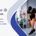
Achilles Tendon Rupture: Surgery vs. Conservative Management — Which Is Better?
An Achilles tendon rupture is one of the most dramatic injuries in sport — a sudden, forceful snap at the back of the ankle that leaves an athlete unable to push off or bear weight. The question that follows almost immediately is: do I need surgery?
In short: the honest answer is that for most active adults and athletes, surgery and well-structured functional rehabilitation produce comparable outcomes — but the right choice depends heavily on age, activity level, rupture characteristics, and how quickly rehabilitation begins. There is no single correct answer for everyone.
At Sancheti Hospital, Pune, our foot, ankle, and sports medicine specialists guide patients through this exact decision daily. This blog lays out the evidence on both sides, clearly and without bias, so you can make an informed decision with your surgeon.
What Is the Achilles Tendon and Why Does It Rupture?
The Achilles tendon is the thickest and strongest tendon in the body, connecting the gastrocnemius and soleus muscles of the calf to the calcaneus (heel bone). It transmits the force of calf contraction into plantarflexion — the push-off that propels every step, jump, and sprint.
Despite its strength, the Achilles is vulnerable to rupture because of a relatively avascular zone approximately 2–6 cm above its heel attachment — a region with poor blood supply that accumulates degenerative change over time. Most ruptures occur here, through tendon tissue that has been silently weakening for years before the final snap.
Common rupture mechanisms:
- Sudden push-off during sprinting, jumping, or change of direction
- Unexpected dorsiflexion (ankle bending upward) against a planted foot — the classic “stepping in a hole” mechanism
- A direct blow to a taut tendon (less common)
The typical patient is a male aged 30–50 — the so-called “weekend warrior” — who is recreationally active but not in structured athletic conditioning. Background Achilles tendinitis or tendinopathy is a significant risk factor, as the degenerative tissue has lower tensile strength than healthy tendon.
Recognising an Achilles Tendon Rupture
The presentation is usually unmistakable:
- A sudden, sharp pain at the back of the ankle — often described as feeling like being “kicked” or “shot” in the heel
- An audible pop at the moment of injury
- Immediate inability to push off on the affected foot
- Visible gap or “dent” in the tendon approximately 2–6 cm above the heel
- Significant swelling and bruising developing over hours
The Thompson test (squeezing the calf with the patient prone — absence of plantarflexion confirms rupture) is the most reliable clinical test and is positive in nearly all complete Achilles tendon ruptures.
Ultrasound confirms the diagnosis, quantifies gap size between the torn ends, and assesses whether the ends approximate in plantarflexion — information that directly influences treatment planning. MRI provides additional detail on tendon quality and partial tear extent when the diagnosis is uncertain.
Surgery vs. Conservative Management: What the Evidence Says
This is the core question — and the evidence has shifted significantly over the past decade.
| Factor | Surgical Repair | Functional Conservative |
| Re-rupture rate | ~3–5% | ~8–12% (with functional rehab) |
| Return to sport | Faster by 4–6 weeks on average | Comparable at 9–12 months |
| Calf strength recovery | Slightly better at 1 year | Comparable by 2 years |
| Complication risk | Wound infection, sural nerve injury, DVT | DVT, skin breakdown (in cast immobilisation) |
| Hospital stay | Day surgery | None |
| Cost | Higher | Lower |
| Best suited for | Young athletes, large gap, delayed presentation | Active adults, small gap, early presentation |
The key finding from multiple randomised controlled trials — including the landmark UKSTAR trial — is that functional rehabilitation with early weight-bearing produces outcomes comparable to surgery in terms of re-rupture rate, strength, and return to activity, provided the rehabilitation protocol is structured and begins promptly.
The earlier view that surgery was always superior has been substantially revised. However, surgery still has a clear role in specific circumstances — and the decision is never one-size-fits-all.
Who Should Choose Surgery?
Surgical repair is generally recommended when:
- Young, competitive athletes requiring the fastest possible return to high-level sport
- Gap >5 mm on ultrasound in plantarflexion — the tendon ends do not approximate adequately for non-surgical healing
- Delayed presentation (>2 weeks) — the tendon ends retract and scar, making functional healing increasingly unlikely without surgical reapproximation
- Re-rupture of a previously conservatively managed tendon
- Associated injuries — for example, combined ankle tendon tears requiring surgical intervention in the same setting
Surgical technique:
Open repair involves a longitudinal incision behind the ankle, direct visualisation of the torn tendon ends, and suture repair — most commonly using a Krackow or modified Kessler technique. Minimally invasive percutaneous repair, using small stab incisions and a specialised device to pass sutures without a large wound, is increasingly used to reduce wound complication rates while preserving the strength advantages of surgical reattachment.
Both approaches are performed under regional or general anaesthesia as day-care procedures at our foot and ankle surgery department at Sancheti Hospital.
Who Can Choose Conservative Management?
Functional conservative management is appropriate when:
- Active adults over 45 with moderate activity demands where the marginal strength advantage of surgery is not functionally meaningful
- Tendon ends approximate on ultrasound in plantarflexion — confirming adequate contact for biological healing
- Early presentation (within 48–72 hours) — before significant retraction occurs
- Medical comorbidities that increase surgical risk — diabetes, peripheral vascular disease, immunosuppression, or medications like fluoroquinolones or steroids (which impair tendon healing)
- Patient preference after informed discussion of both options
What conservative management actually involves:
Conservative does NOT mean simply resting in a cast. The evidence for conservative management comes from functional rehabilitation protocols — not traditional immobilisation. The key steps are:
- Immediate immobilisation in a walking boot with heel raises (plantarflexion) to approximate the tendon ends — within hours of injury
- Early weight-bearing at 2 weeks in the boot — critical; delayed weight-bearing significantly increases re-rupture risk
- Progressive reduction of heel raises over weeks 2–8 as tendon healing progresses
- Formal physiotherapy beginning at week 2 — range of motion, calf activation, proprioception
- Boot weaned at weeks 8–10 as the tendon is confirmed healed on ultrasound
The distinction between good and poor conservative outcomes is almost entirely explained by whether the protocol was truly functional (early weight-bearing, structured rehabilitation) or traditional (prolonged cast immobilisation with late mobilisation). The latter carries re-rupture rates exceeding 20% — unacceptably high and the source of much of the historical bias toward surgery.
Rehabilitation: Phase That Determines the Outcome
Regardless of whether surgery or conservative management is chosen, rehabilitation quality drives the outcome. Both pathways follow a broadly similar staged progression:
| Phase | Timeline | Key Goals |
| Protection | Weeks 1–6 | Tendon healing, protected weight-bearing in boot, early calf activation |
| Mobilisation | Weeks 6–10 | Boot weaned, full weight-bearing, range-of-motion restoration |
| Strengthening | Weeks 10–20 | Progressive calf raises, single-leg heel raise target |
| Power & sport-specific | Months 5–7 | Plyometrics, running reintroduced, sport-specific drills |
| Return to sport | Months 8–12 | Full competitive return with strength symmetry confirmed |
The single-leg heel raise is the functional benchmark of Achilles rehabilitation — the ability to perform 25 consecutive single-leg heel raises on the injured side, equal in height and control to the uninjured side, before return to running is cleared.
A structured physiotherapy and rehabilitation program that progresses each phase based on objective milestones — not calendar time — is the single most important determinant of outcome in both surgical and conservative management.
Complications: What to Watch For
Surgical complications:
- Wound infection — the most common; risk is higher in diabetics and smokers
- Sural nerve injury — temporary or permanent numbness along the outer foot
- DVT — risk is present in any lower limb surgery and is mitigated with anticoagulation and early mobilisation
- Tendon adhesions — stiffness from scar tissue formation, addressed with physiotherapy
Conservative complications:
- Re-rupture — the primary risk; highest if weight-bearing is delayed or rehabilitation is inadequate
- Tendon elongation — healing in a lengthened position reduces calf strength and plantarflexion power; prevented by maintaining heel raise position during early healing
- DVT — present in conservative management too, particularly with prolonged immobilisation
Our sports medicine specialists at Sancheti Hospital monitor healing progress with serial ultrasound at key time points — confirming tendon continuity and approximation before advancing the rehabilitation protocol — for both surgical and conservatively managed patients.
Making the Decision: A Practical Framework
| Patient Profile | Recommended Approach |
| Competitive athlete under 40, wants fastest return | Surgery |
| Active adult, early presentation, tendon ends approximate | Functional conservative |
| Delayed presentation (>2 weeks) | Surgery |
| Medical comorbidities increasing surgical risk | Conservative |
| Re-rupture after previous conservative management | Surgery |
| Older adult (>60) with low activity demands | Conservative |
| Patient unwilling to accept 8–12% re-rupture risk | Surgery |
No framework replaces an individualised discussion with your surgeon — but this table reflects how most experienced foot and ankle specialists approach the decision at Sancheti Hospital.
Key Takeaways
- Achilles tendon ruptures most commonly occur in the avascular zone 2–6 cm above the heel, in degeneratively weakened tendon tissue, during sudden push-off or unexpected dorsiflexion.
- The Thompson test is the most reliable clinical sign; ultrasound confirms the diagnosis and guides the treatment decision.
- Surgery and functional conservative management produce comparable outcomes in re-rupture rate and long-term function — provided conservative management uses an early weight-bearing protocol, not prolonged cast immobilisation.
- Surgery offers a modest advantage in faster return to sport and lower re-rupture risk (3–5% vs 8–12%) and is preferred for competitive athletes, delayed presentations, and large tendon gaps.
- Conservative management is a valid, evidence-supported choice for active adults with early presentation, good tendon approximation, and no surgical risk factors.
- Rehabilitation quality is the common factor that determines outcome in both pathways — the single-leg heel raise is the key functional milestone before return to sport is cleared.
- At Sancheti Hospital, Pune, our foot, ankle, and sports medicine specialists make this decision jointly with each patient — using ultrasound assessment, activity profiling, and an honest presentation of both options — rather than defaulting to a single approach for everyone.
Frequently Asked Questions (FAQs)
Q1. How do I know if my Achilles tendon is fully ruptured or just partially torn?
A complete rupture typically produces an immediate inability to push off, a palpable gap in the tendon, and a positive Thompson test. A partial tear causes significant pain and weakness but partial push-off is usually preserved and the Thompson test is negative or equivocal. Ultrasound confirms the distinction clearly and also quantifies how much of the tendon is involved — information that directly influences whether surgery, conservative management, or a period of observation is appropriate.
Q2. Can I walk after an Achilles tendon rupture?
Many patients can hobble on a ruptured Achilles using the toe flexors and peroneals for limited propulsion, which leads to the common misdiagnosis of “sprain.” The inability to perform a single-leg heel raise — not the ability to walk — is the key functional indicator of a complete rupture. Walking capacity alone should never be used to exclude the diagnosis.
Q3. What happens if an Achilles tendon rupture is not treated at all?
Untreated complete ruptures heal in a lengthened, disorganised scar tissue bridge that significantly reduces calf power and plantarflexion strength — often permanently. The patient can usually walk, but running, jumping, and stair climbing are severely impaired. Surgical repair of a chronically neglected rupture is significantly more complex than acute repair, often requiring tendon transfer procedures (such as flexor hallucis longus transfer) to bridge the gap.
Q4. Is the Achilles tendon as strong after repair as it was before the rupture?
With well-executed surgical repair and complete rehabilitation, the repaired tendon recovers most of its functional strength. Studies show calf strength at 2 years post-repair is typically 85–95% of the uninjured side. Full strength symmetry is achievable in most patients but requires consistent completion of the rehabilitation program — particularly the progressive calf loading phases.
Q5. Can Achilles tendon rupture be prevented?
The risk can be meaningfully reduced through structured calf and Achilles conditioning, gradual training load increases, avoiding sudden return to high-intensity sport after periods of inactivity, and addressing Achilles tendinopathy promptly before degenerative change accumulates. Fluoroquinolone antibiotics and corticosteroid injections into the tendon are known risk factors for rupture and should be used with caution in active individuals with known tendon pathology.